Brain Reprogramming: Scientists Manage to Stimulate Memory Without Touching The Deep Brain

This study showed that it is possible to stimulate a deep region of the brain, responsible for memory, using magnetic pulses applied to the surface of the head. By choosing specific points in the brain that are connected to this region, the researchers were able to activate it indirectly. The results indicate that the stronger this connection in each person, the better the effect of the stimulation. This discovery paves the way for non-invasive and personalized treatments for memory and mental health problems.
Memory is one of the most important functions of the human brain, and one of the main structures responsible for it is the hippocampus, a region located deep in the brain. In addition to helping form and retrieve memories, the hippocampus also participates in processes such as learning and spatial orientation. Therefore, scientists have been searching for ways to stimulate this region to improve memory and treat diseases such as Alzheimer's and depression.
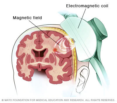
One of these approaches is transcranial magnetic stimulation, a non-invasive technique that uses magnetic pulses applied to the surface of the head to influence brain activity.
However, there is a significant challenge: the hippocampus is located deep within the brain, making it difficult to reach directly with this technique. The solution explored in this study was to stimulate a more superficial region of the brain, the parietal cortex, which has natural connections with the hippocampus.
The idea is similar to flipping a distant switch that, because it is connected by internal wires, can activate another, deeper point. Thus, the researchers investigated whether it would be possible to influence the hippocampus indirectly by stimulating brain regions that "communicate" with it.
To test this accurately, the scientists used a personalized approach. First, they mapped each participant's brain to identify which areas of the parietal cortex had the strongest functional connection to the hippocampus. This functional connection means that these regions tend to work together, activating in a coordinated way.
This mapping was done using brain imaging scans, allowing them to choose specific points to stimulate in each person, instead of using a standard location that was the same for everyone.
The most innovative part of the study was the combination of different techniques to observe what was happening inside the brain. In eight patients who had already undergone neurological surgery and had electrodes implanted in their brains, the researchers applied magnetic stimulation externally and, at the same time, directly recorded the activity of the hippocampus from within.
This is rare, as it is not normally possible to measure the activity of deep regions with such precision in humans. This approach allowed them to verify in real time whether the stimulus actually reached the hippocampus.

Furthermore, the scientists also studied a larger group of seventy-nine healthy people using imaging scans that measure brain activity. In these participants, they observed how the brain responded to stimulation over time. This helped confirm whether the effects seen in patients with electrodes also occurred in people without neurological conditions, increasing confidence in the results.
The results showed that the stimulation worked like a “remote control”: by stimulating the correct parietal cortex, the hippocampus was activated with specific patterns of activity. However, this effect varied from person to person. The stronger the connection between the two regions in an individual's brain, the greater the hippocampal response. This reinforces the importance of personalization, since using a generic stimulation point did not produce the same effect.

Another important finding was that, when stimulation was applied repeatedly, it could modify certain natural rhythms of the hippocampus, especially those linked to memory. These rhythms are like "waves" of brain activity that help organize information. Altering them can directly influence the ability to learn and remember.
Taken together, this study demonstrates that it is possible to influence deep brain regions indirectly, safely, and in a personalized way, without the need for surgery. This paves the way for the development of new treatments for diseases that affect memory and emotional functioning.
In the future, techniques like this could be used to "reset" altered brain circuits, offering a promising alternative to improve the quality of life for many people.
READ MORE:
Multimodal evidence for hippocampal engagement and modulation by functional connectivity-guided parietal TMS
Zhuoran Li, Nicholas T. Trapp, Joel Bruss, Xianqing Liu, Kang Wu, Ziyan Chen, Amit Etkin, Matthew A. Howard, Aaron D. Boes, and Jing Jiang
Nature Communications
DOI:10.1038/s41467-026-70346-x
Abstract:
Hippocampal activity supports memory and many other brain functions. Transcranial magnetic stimulation (TMS) guided by hippocampal functional connectivity (FC) shows promise in improving memory, but direct neural evidence of its capacity to engage and modulate hippocampal activity is lacking. Here we combined TMS with intracranial electroencephalography (iEEG) in 8 neurosurgical patients and with functional magnetic resonance imaging (fMRI) in 79 neurologically healthy participants. We identified that (1) single-pulse TMS to individualized parietal cortex guided by hippocampal-FC preferentially evoked distinct temporal and spectral activity patterns in the hippocampus, (2) variability in TMS-evoked hippocampal responses related to individual differences in parietal-hippocampus FC strength, and (3) repetitive TMS to hippocampal-FC-guided parietal cortex selectively suppressed hippocampal theta oscillations. These findings provide multimodal causal neural evidence and important mechanistic insights supporting the development of personalized neuromodulation strategies aimed at improving hippocampus-dependent functions.


Comments